Digital Imaging & Diagnostic Technologies
Advanced Digital Imaging in Sarasota for Precise Diagnosis and Treatment Planning
Cutting-Edge Technologies to Enhance Accuracy and Improve Your Dental Care Outcomes

Advanced Dental X-Rays for Superior Diagnosis and Care
Dental radiographs are an essential part of your overall dental health evaluation. They are especially effective for detecting tooth decay and gum disease by assessing bone levels around teeth and dental implants. Digital dental X-rays offer several advantages over traditional film X-rays, including instant image viewing, enhanced resolution and detail, and reduced radiation exposure. Your dental X-rays are downloaded onto a computer, allowing us to use advanced software to enhance the images for improved diagnosis and treatment planning.
At Misch Implant & Aesthetic Dentistry, we utilize the award-winning DEXIS imaging system. DEXIS uses Advanced CleanCapture technology to enhance detail and produce high-resolution images. The system processes images with ClearVu™, resulting in clear, highly detailed radiographs.
We also use a handheld X-ray machine called the NOMAD Pro 2, which gives our team greater flexibility and easier positioning of the X-ray unit to capture better images. It also allows our staff to remain in the room with the patient, helping to keep them calm and properly positioned for a more positive and comfortable experience.
Revolutionizing Implant Dentistry with Advanced 3D Imaging Technology
One of the most significant advances in implant dentistry over the past 20 years has been the ability to obtain three-dimensional radiographs of the jawbone in-office to assess anatomy and bone volume for implant placement. Cone beam computed tomography (CBCT) is a variation of traditional CT scan systems. By using advanced computer technology and software, CBCT requires significantly less radiation to produce 3D images compared to the CT scan units used in medicine.
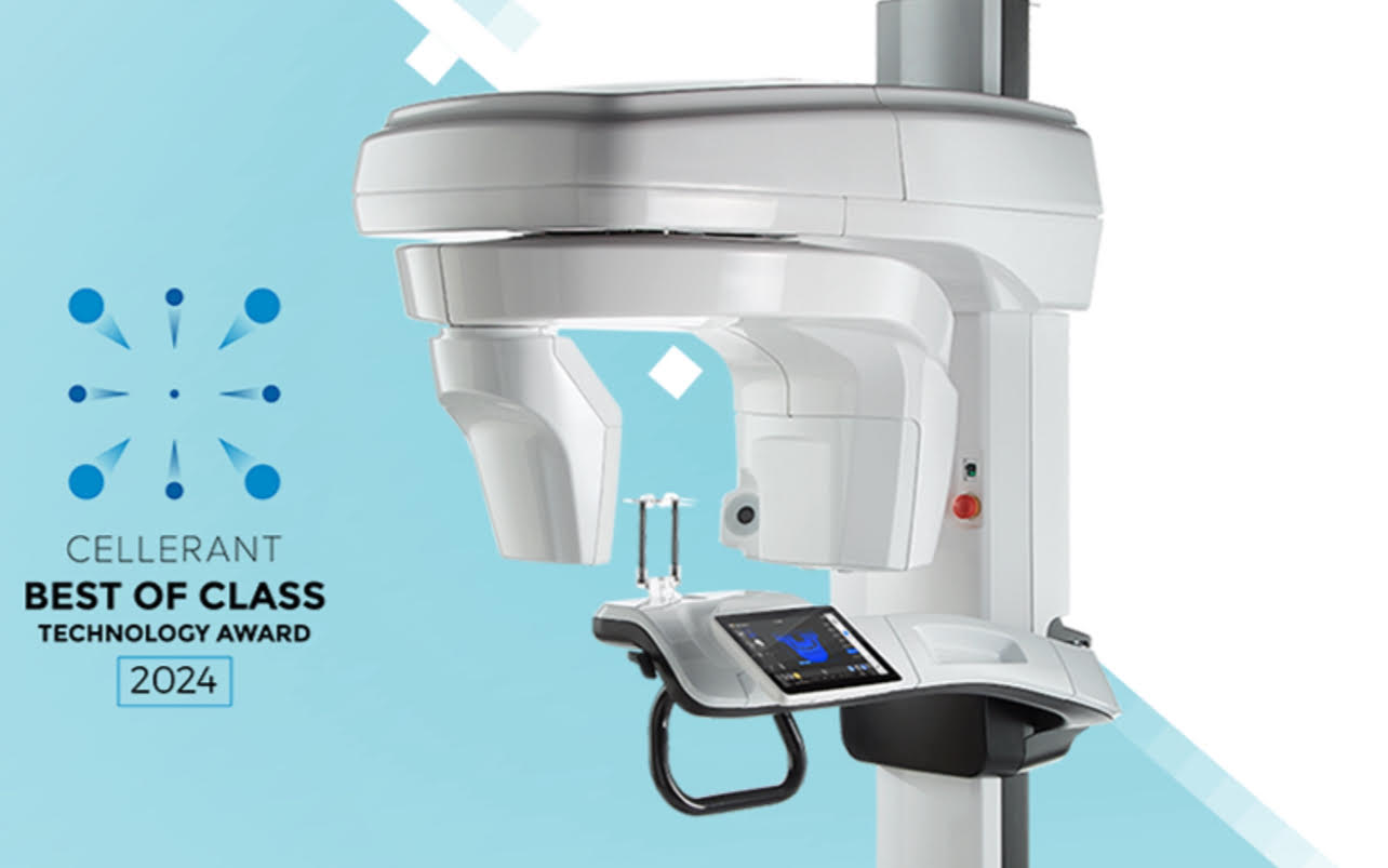
We have recently acquired the next generation of cone beam CT scanners: the Carestream 9600. This unit has received the Cellerant “Best of Class” award in dental technology. It utilizes artificial intelligence to deliver more accurate scans on the first attempt. Its high-resolution imaging surpasses that of other units on the market, enhancing our diagnostic capabilities and improving surgical precision.
This new scanner also features an innovative Face Scan function that generates realistic 3D facial images, which are superimposed over the underlying facial bones. This provides additional diagnostic information to help us plan your case for the best possible outcome. The scanner includes a low-dose mode to further minimize radiation exposure compared to other CT scanners.
The virtual dental implant surgery feature has also been optimized, enabling our board-certified surgeons to digitally plan your bone graft or dental implant procedure in advance. Using your CT scan images, we can select the most effective approach and materials for your specific case. It’s like rehearsing your surgery before the real one—because champions win by preparing for success.

Photogrammetry in Implant Dentistry: Achieving Precision and Efficiency in Implant Restorations
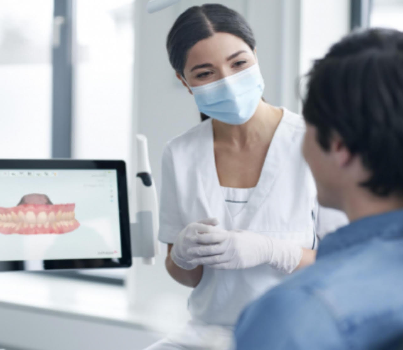
Photogrammetry is a new technique used in implant dentistry to capture images and create highly accurate 3D models of the implant positions. This technique is more precise than traditional impressions. It involves attaching photogrammetry-coded abutments to implants and capturing multiple images from different angles using a specialized camera. Using software the images are stitched together to create a 3D representation.
Once the photogrammetry scan is complete, a digital file extension (STL or XML file) is created that contains all the interrelated information on implant geometries and interfaces. This data can then be uploaded to computer-aided design software to allow for digital alignment pairing and designing of the prosthesis. After the prosthesis is designed, an STL file can be sent to a 3D printer. The prosthesis can then be 3D printed at our office and delivered to the patient the same day.
The benefits of photogrammetry are numerous. This is a much better experience for the patient compared to using impression material and trays in the mouth. This technique offers increased accuracy which is crucial for fabricating precise implant restorations. In full-arch implant dentistry, obtaining an accurate representation of the implant positions is imperative to the fit and long-term success of the prosthesis. It can also reduce the number of visits required for full-arch implant prostheses. The high precision of photogrammetry contributes to more predictable and successful restorative outcomes.
FAQs
Photogrammetry involves capturing detailed 3D images of the mouth using specialized cameras and software. This technology allows for precise planning of implant placement, ensuring better fit and function of the final restoration. In Sarasota, some dental practices utilize photogrammetry to enhance the accuracy and outcomes of implant procedures.
Digital radiographs are a modern form of dental X-rays that use electronic sensors to capture images of the teeth and bones. Compared to traditional film X-rays, digital radiographs offer several advantages:
- Reduced Radiation Exposure: Digital sensors require less radiation to produce high-quality images.
- Immediate Image Availability: Images are available instantly, allowing for quicker diagnosis and treatment planning.
- Enhanced Image Quality: Digital images can be adjusted for better clarity and detail.
- Environmentally Friendly: Eliminates the need for chemical processing associated with traditional film.
Cone beam computed tomography (CBCT) is an advanced imaging technique that provides three-dimensional images of the teeth, jaw, and surrounding structures. Unlike traditional X-rays, CBCT scans offer detailed views that assist in:
- Implant Planning: Assessing bone density and structure for optimal implant placement.
- Endodontic Evaluations: Identifying root canal anatomy and potential complications.
- Orthodontic Assessments: Evaluating tooth alignment and jaw relationships.
- TMJ Analysis: Examining the temporomandibular joint for disorders.
CBCT scans are particularly beneficial when traditional X-rays do not provide sufficient information.
Photogrammetry is a technique that uses high-resolution cameras to capture detailed images of the oral cavity from multiple angles. These images are then processed to create accurate 3D models of the patient’s dental structures. In implant dentistry, photogrammetry offers several benefits:
- Enhanced Accuracy: Provides precise measurements for implant placement.
- Improved Fit: Ensures better alignment and integration of dental prosthetics.
- Reduced Need for Traditional Impressions: Minimizes patient discomfort and the potential for inaccuracies.
- Streamlined Workflow: Facilitates quicker turnaround times for restorations.
This technology is especially useful in full-arch implant cases, where precision is critical.
Yes, advanced imaging technologies like digital radiographs, CBCT, and photogrammetry are designed with patient safety in mind. Digital radiographs and CBCT scans use minimal radiation, adhering to the ALARA (As Low As Reasonably Achievable) principle to minimize exposure. Additionally, photogrammetry is a non-invasive technique that does not involve radiation. Dental professionals in Sarasota ensure that these technologies are used appropriately and safely to enhance patient care.
Just Call - Booking Is That Easy!
Give Us a Call
Tell Us What You Need
Pick a Time That Works for You

